Chapter 1- Organization of the Body
For the first chapter of Anatomy and Physiology we studied the organization of the body. The body is organized into many different categories. The first way the body is categorized is by level of organization. The levels are:
1.Chemical
2.Cellular
3. Tissue
4. Organ
5. System
The next way the body is categorized is by body planes. The 3 planes are:
1. Sagittal- divides the body into right and left sections
2. Transverse- (aka horizontal) divides the body into top and bottom sections
3. Coronal- (aka vertical) divides the body into front and back sections
The next way the body is divided is by cavity.
1. Dorsal Cavity (back)
a. cranial cavity
b.spinal cavity
2. Ventral Cavity (front)
a. thoracic (chest) cavity
b. abdminopelvic (belly) cavity- the picture above depicts the subdivisions of the abdminopelvic region.
In this chapter we also learned directional terms such as:
anterior- in front of
posterior- in back of
medial- toward midline
lateral- away from midline
superior- above
inferior- below
superficial- nearer to surface
deep- further from surface
proximal- nearer to the torso
distal- further from torso
The Skeletal System
Our next chapter for anatomy focuses on the human skeleton.The human skeleton is composed of 300 bones at birth and by the time adulthood is reached, some bones have fused together to give a total of 206 bones in the body. The bone mass in the skeleton reaches maximum density around age 30. The human skeleton can be divided into the axial skeleton and the appendicular skeleton. The axial skeleton is formed by the vertebral column, the rib cage and the skull. The appendicular skeleton, which is attached to the axial skeleton, is formed by the pectoral girdles, the pelvic girdle and the bones of the upper and lower limbs.The human skeleton serves six major functions; support, movement, protection, production of blood cells, storage of ions and endocrine regulation.
The Muscular System
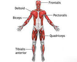
The muscular system is an organ system consisting of skeletal, smooth and cardiac muscles. It permits movement of the body, maintains posture, and circulates blood throughout the body. The muscular system in vertebrates is controlled through the nervous system, although some muscles (such as the cardiac muscle) can be completely autonomous. Together with the skeletal system it forms the musculoskeletal system, which is responsible for movement of the human body.
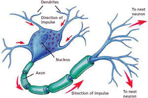
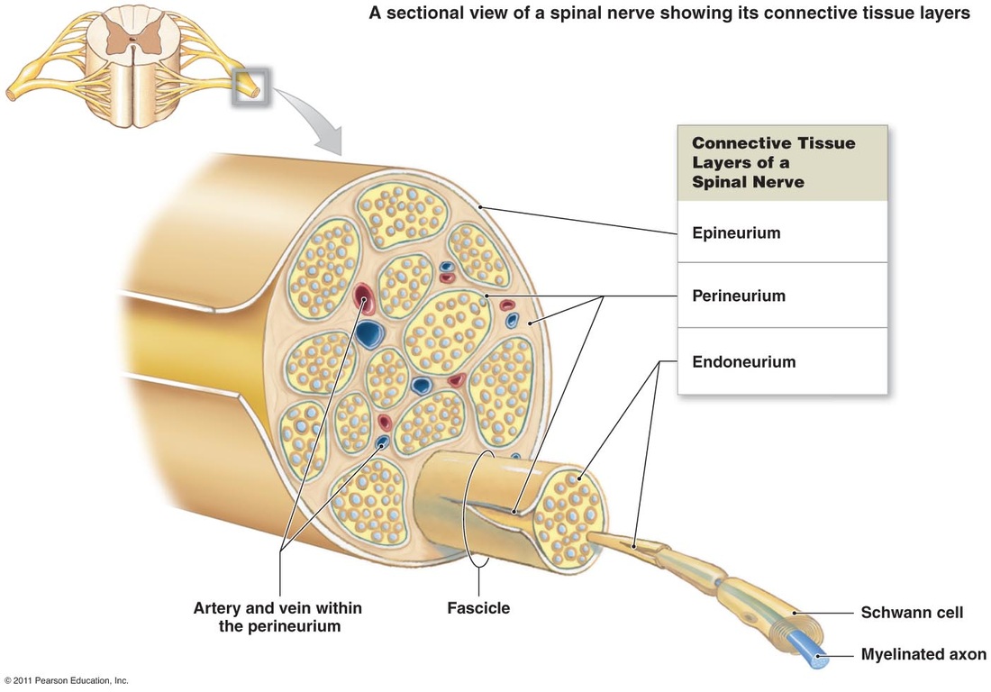
Nervous System
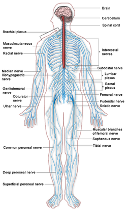
The nervous system is a complex collection of nerves and specialized cells known as neurons that transmit signals between different parts of the body. Vertebrates — animals with backbones and spinal columns — have central and peripheral nervous systems.
The central nervous system is made up of the brain, spinal cord and retina. The peripheral nervous system consists of sensory neurons, ganglia (clusters of neurons) and nerves that connect to one another and to the central nervous system.
From a top view, notice how the brain is divided into two halves, calledhemispheres. Each hemisphere communicates with the other through the corpus callosum, a bundle of nerve fibers. (Another smaller fiber bundle that connects the two hemispheres is called the anterior commissure).
Some differences between the peripheral nervous system (PNS) and the central nervous system (CNS):
- In the CNS, collections of neurons are called nuclei. In the PNS, collections of neurons are called ganglia.
- In the CNS, collections of axons are called tracts. In the PNS, collections of axons are called nerves.
- Sensory (afferent) - carry information INTO the central nervous system from sense organs or motor (efferent) - carry information away from the central nervous system (for muscle control).
- Cranial - connects the brain with the periphery or spinal - connects the spinal cord with the periphery.
- Somatic - connects the skin or muscle with the central nervous system or visceral - connects the internal organs with the central nervous system.

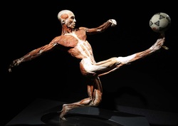
Body World Field Trip
Our next big thing in Anatomy was a class field trip to Body World Vital in Boston. Body world is an exhibit that uses human donor bodies to help show the function and structure of the inside of the human body. These detailed anatomical studies, compositions, and representations allow visitors a penetrating gaze at what lies beneath the skin--the keenly intelligent design and function of the human body. In this collaboration between donors, anatomist, and visitors, the donors – having given legal consent during their lifetimes to participate in Body world– act as guides and teachers, across time, on this motivational journey towards health and vitality. Physician and anatomist Gunther von Hagens’ Body world exhibitions are the precedent-setting museum anatomical exhibitions of real human donor bodies. The bodies are preserved through a remarkable process called Plastination, invented by Dr. von Hagens, Plastination is a method of extracting bodily fluids and fat from specimens, and replacing them with reactive resins and polymers, enabling visitors to actually see inside the human body, learn how it works, and how it can be affected by disease.
